

Dr. Sunil Chopra
Radiologist & Nuclear Medical Physician
MBBS (Delhi), M. Rad (UM), F’ship in Nuc Med Imaging (IAEA)
Clinical Indications
- Detection of skeletal metastases for Initial Staging, Disease extent and assess Response to treatment
- Stress injuries and fractures
- Bone viability after surgery or trauma
- Complications of joint prostheses, bone graft viability, fracture nonunion
- Osteonecrosis of femoral head and mandible
- Metabolic bone diseases like Osteoporosis, Renal osteodystrophy, Paget`s disease
- Osteomyelitis
Bones not only support the body structure, but are also important in producing blood cells and storing minerals. Diseases or conditions which involve the bones such as tumours, degenerative diseases, infections and injuries can therefore affect these functions.
Various imaging techniques can be used for imaging bones, including X-rays, CT scans and MRI. Besides these, nuclear medicine imaging using radioactive isotopes such as Technetium have been used for many years. With the invention of the PET-CT scanner, combining both PET and CT technology in a single scanner, new and more advanced imaging techniques are now available toassess bony involvement in diseases.
Sodium Fluoride PET-CT (NaF PET-CT) is a new imaging service offered at Loh Guan Lye Specialists Centre from July 2016, providing significantly improved quality and definition compared to other nuclear medicine bone imaging techniques.
The most common indication for the NaF PET-CT scan is in various cancers that may involve the bones, not only to detect spread of cancer to the bones, but also to assess the degree of bony involvement as well as to monitor the response to treatment.
Other indications include the detection of stress injuries and fractures as well as assessment of bone viability after surgery and also to detect complications related to bone implants.
Various clinical indications for NaF PET-CT Scan
This scan uses radioactive Fluoride bonded to sodium, which is then injected into the body and is taken up by areas of increased bone activity. Images of the whole body are then obtained on a PETCT scanner after 45-60 minutes, providing a detailed map of normal and abnormal bone activity in various conditions.


Compared to other bone scanning techniques using radioactive Technetium, NaF PET-CT is more sensitive and specific in detecting bony lesions (tumour involvement of the bones). Not only does NaF PET-CT detect up to 45 % more lesions, it has other advantages over older bone scanning methods as well. The preparation for this scan is less inconvenient for patients, with no specific restrictions related to diet, medications and lifestyle.
The waiting time and scanning time is significantly less, while image quality is significantly better than older bone scanning techniques. Fusion of PET images with CT images allows precise localisation of lesions and better assessment without the extra expense and inconvenience of having to perform another examination to assess bone lesions found on the Technetium scans.
Interpretation of PET-CT images is done by trained specialists working on dedicated workstations to assess the images. Other clinical and investigative findings are taken into account when reporting the scans to obtain as much information as possible in order to provide the best treatment to the patient.
Loh Guan Lye Specialists Centre is proud to be the first centre in the country to provide NaF PET-CT imaging services, consistent with our commitment to providing the best treatment using the latest technology.


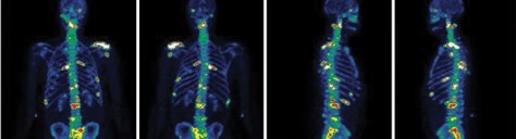
Comparison between conventional bone scan using Technetium (left) and NaF PET-CT (right) in two patients with tumour involving the bones showing significantly better image quality of the NaF PETCT scan with more areas of bone involvement seen compared to the Technetium scan.

